Cortical tension regulates desmosomal morphogenesis
Moch M, Schieren J and Leube RE
Mechanical stability is a fundamental and essential property of epithelial cell sheets. It is in large part determined by cell-cell adhesion sites that are tightly integrated by the cortical cytoskeleton. An intimate crosstalk between the adherens junction-associated contractile actomyosin system and the desmosome-anchored keratin intermediate filament system is decisive for dynamic regulation of epithelial mechanics. A major question in the field is whether and in which way mechanical stress affects junctional plasticity. This is especially true for the desmosome-keratin scaffold whose role in force-sensing is virtually unknown. To examine this question, we inactivated the actomyosin system in human keratinocytes (HaCaT) and canine kidney cells (MDCK) and monitored changes in desmosomal protein turnover.
Partial inhibition of myosin II by para-nitro-blebbistatin led to a decrease of the cells' elastic modulus and to reduced desmosomal protein turnover in regions where nascent desmosomes are formed and, to a lower degree, in regions where larger, more mature desmosomes are present. Interestingly, desmosomal proteins are affected differently: a significant decrease in turnover was observed for the desmosomal plaque protein desmoplakin I (DspI), which links keratin filaments to the desmosomal core, and the transmembrane cadherin desmoglein 2 (Dsg2). On the other hand, the turnover of another type of desmosomal cadherin, desmocollin 2 (Dsc2), was not significantly altered under the tested conditions. Similarly, the turnover of the adherens junction-associated E-cadherin was not affected by the low doses of para-nitro-blebbistatin. Inhibition of actin polymerization by low dose latrunculin B treatment and of ROCK-driven actomyosin contractility by Y-27632 treatment also induced a significant decrease in desmosomal DspI turnover. Taken together, we conclude that changes in the cortical force balance affect desmosome formation and growth. Furthermore, they differentially modulate desmosomal protein turnover resulting in changes of desmosome composition. We take the observations as evidence for a hitherto unknown desmosomal mechanosensing and mechanoresponse pathway responding to an altered force balance.
High-dose para-nitro-blebbistatin obscures Dsg-2-mCerulean but not DspI-mApple fluorescence. The time-lapse recording shows maximum intensity projections of the fluorescence in the 5 lower focal planes of HaCaT keratinocytes co-expressing DspImApple (left) and Dsg2-mCerulean (middle; merged images at right). Note the well-delineated
co-distribution of DspI-mApple and Dsg2-mCerulean during the 15 min prior to the addition of 20 μM para-nitro-blebbistatin, which is difficult to track afterwards because of the strong autofluorescence of the drug, which is detectable in the cells within 2 min and subsequently accumulates to levels above signal saturation.
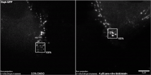
Desmosomal protein turnover can be measured in FRAP experiments under different conditions. The two time-lapse fluorescence series show fluorescence recovery of fluorescently tagged DspI (DspI-GFP) in HaCaT keratinocytes after addition of 0.5% DMSO alone (left) and after addition of 0.5% DMSO with 4 μM para-nitroblebbistatin (right). The movies show sum-projections of the recorded fluorescence in selected regions as described in Fig. 3C. The first frame presents the cells 60 s before bleaching was initiated and the second frame shows the situation directly after bleaching (which took 7-10 s). Fluorescence recovery measurements were started one minute after bleaching (frame 3, t=0) and
fluorescence intensity was set as 0%. In this way, diffusion-related rapid fluorescence recovery was not taken into account for the quantitative measurements, which are shown in the subsequent
frames for 14 minutes. Note that the cells have neighbors on the left, right and top, but not at the
bottom.

DspI-GFP clustering at nascent desmosomes continues in the presence of low-dose para-nitro-blebbistatin. The time-lapse recording shows the projected 7 lower planes of DspI-GFP fluorescence in a transfected HaCaT keratinocyte next to nontransfected keratinocytes. The imaged region shows cells in the periphery of a cell colony (to the
right as illustrated in Fig. S3). Note that new DspI-positive puncta appear at the upper and lower left.
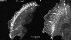
Low dose latrunculin B and para-nitro-blebbistatin treatment affect the actin cytoskeleton differently. The time-lapse recordings of the projected lower 5
focal planes show Actin-mApple fluorescence in HaCaT keratinocytes 20 min prior to the addition of either 0.2 μM latrunculin B (left) or 4 μM para-nitro-blebbistatin (right). Note the partial restructuring of the actin cytoskeleton during the ensuing 40 min recording, which is different in both situations. Latrunculin B treatment partially disrupts the actin network whereas para-nitro-blebbistatin induces a translocation of the network toward the cell center.