Induction of rapid and reversible cytokeratin filament network remodeling by inhibition
Strnad P, Windoffer R, Leube RE, 2002
The cytokeratin filament network is intrinsically dynamic, continuously exchanging subunits over its entire surface, while conferring structural stability on epithelial cells. However, it is not known how cytokeratin filaments are remodeled in situations where the network is temporarily and spatially restricted. Using the tyrosine phosphatase inhibitor orthovanadate we observed rapid and reversible restructuring in living cells, which may provide the basis for such dynamics.
By examining cells stably expressing fluorescent cytokeratin chimeras, we found that cytokeratin filaments were broken down and then formed into granular aggregates within a few minutes of orthovanadate addition. After drug removal, gradual reincorporation of granules into the filament network was observed for aggregates that were either part of residual filaments or stayed in close apposition to remaining filaments. Even when cytokeratin filaments were no longer detectable, granules with low mobility were still able to reestablish a cytokeratin filament network. This process took less than 30 minutes and occurred at multiple foci throughout the cytoplasm without apparent correlation to alterations in the actin- and tubulin-based systems.
Interestingly, the short-lived and rather small orthovanadate-induced cytokeratin granules contained the cytoskeletal crosslinker plectin but lacked the cytokeratin-solubilising 14-3-3 proteins. By contrast, the long-lived and larger cytokeratin aggregates generated after treatment with the serine/threonine phosphatase inhibitor okadaic acid were negative for plectin but positive for 14-3-3 proteins. Taken together, our observations in living orthovanadate-treated interphase cells revealed modes of cytokeratin remodeling that qualify as basic mechanisms capable of rapidly adapting the cytokeratin filament cytoskeleton to specific requirements.
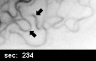
Time-lapse fluorescence microscopy of hepatocellular carcinoma-derived PLC clone PK18-5 stably expressing fluorescent fusion protein HK18-YFP depicting the distribution of labeled CKs. The inverse contrast images were recorded every second and demonstrate details of CKF-disruption starting 234 seconds after addition of OV (50 mM)
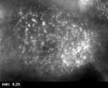
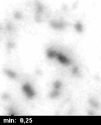
Inverse fluorescence microscopy of living OV-treated (10 mM) AK 13-1 cells detecting HK13-EGFP. Images were recorded every 8 seconds and are presented as averages of consecutive frames. Fine granules are formed within filaments reaching a maximum 12 minutes after OV addition and gradually re-integrate into CKFs with almost no thickenings remaining by 38 minutes (see also Fig. 10).
Time-lapse fluorescence microscopy of AK 13-1 cells monitoring the distribution of fusion protein HK13-EGFP after a 3 minute treatment with 10 mM OV. Pictures were recorded every 15 seconds. A complete CKF network was rebuild within less than 30 minutes with no remaining granules and a very homogenous composition
High magnification of a region taken from movie 3 showing the distribution of HK13-EGFP in AK13-1 cells after a 3 minute treatment with 10 mM OV (recording intervals 15 seconds). For best visualization pictures are shown as averaged consecutive frames. Note the elongation and fusion of granules and their integration into a fine CKF network.