Dissection of keratin dynamics: different contributions of the actin and microtubule systems
Wöll S, Windoffer R, Leube RE, 2005
It has only recently been recognized that intermediate filaments (IFs) and their assembly intermediates are highly motile cytoskeletal components with cell-type- and isotype-specific characteristics. To elucidate the cell-type-independent contribution of actin filaments and microtubules to these motile properties, fluorescent epithelial IF keratin polypeptides were introduced into non-epithelial, adrenal cortex-derived SW13 cells. Time-lapse fluorescence microscopy of stably transfected SW13 cell lines synthesizing fluorescent human keratin 8 and 18 chimeras HK8-CFP and HK18-YFP revealed extended filament networks that are entirely composed of transgene products and exhibit the same dynamic features as keratin systems in epithelial cells.
Detailed analyses identified two distinct types of keratin motility: (I) Slow (approximately 0.23 microm/min), inward-directed, continuous transport of keratin filament precursor particles from the plasma membrane towards the cell interior, which is most pronounced in lamellipodia. (II) Fast (approximately 17 microm/min), bidirectional and intermittent transport of keratin particles in axonal-type cell processes. Disruption of actin filaments inhibited type I motility while type II motility remained. Conversely, microtubule disruption inhibited transport mode II while mode I continued. Combining the two treatments resulted in a complete block of keratin motility. We therefore conclude that keratin motility relies both on intact actin filaments and microtubules and is not dependent on epithelium-specific cellular factors.
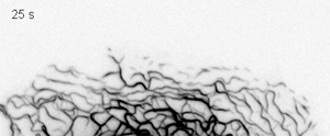
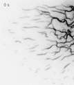
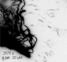
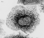
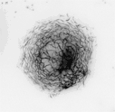
Time-lapse fluorescence microscopy (inverse presentation) at high power of living SK8/18-2 cells detecting dynamics of HK18-YFP distribution in the presence of 30 μM latrunculin. The segment was taken from the same recording that was used to prepare movie 5. Recording intervals, 25 s.