Actin-dependent dynamics of keratin filament precursors
Kölsch A, Windoffer R, Leube RE
Actin filament and microtubule growth characteristics are defined by their different plus and minus ends. In contrast, intermediate filaments lack this type of polarity. Yet, intermediate filament network growth occurs by selective addition of newly formed and polymerizing keratin particles at peripheral network domains thereby allowing polarized network reorganization. To examine this process at high resolution in living cells, mammary epithelium-derived, immortalized EpH4-cells were infected with retroviral cDNA constructs coding for human keratin 18-fluorescent protein hybrids.
Several stable cell lines were established presenting characteristic fluorescent keratin filament (KF) networks. These cells contain particularly large and abundant lamellipodia in which nascent keratin particle dynamics are easily detected by time-lapse fluorescence microscopy. These keratin particles originate close to the plasma membrane, translocate continuously toward the cell center, and integrate end-on into the peripheral KF network. We show that this inward-directed transport relies on intact actin filaments. After treatment with the actin filament-disrupting drug cytochalasin newly polymerizing keratin assemblies still appear in the peripheral cytoplasm but remain stationary. On the other hand, nocodazole-mediated disruption of microtubules does not affect the centripetal KF precursor transport.
From these and other observations a model is deduced which postulates that focal adhesion-dependent keratin polymerization occurs in forming lamellipodia and that transport of newly formed keratin particles is mediated by actin filaments until network integration. This mechanism allows extension of the KF network toward the leading edge in migrating cells and may be of relevance for tissue development and regeneration.
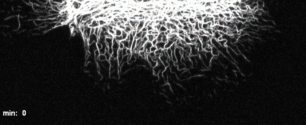
KFP-formation in the periphery of EK18-1-cells
Time-lapse fluorescence recording (projection of 13 focal image planes) of an EK18-1 cell producing fluorescent HK18-YPF protein chimera. Note the abundant amount of emerging KFPs in the cell periphery, which are transported towards the cell centre. The display rate is 10 frames/s, and images were taken every minute.
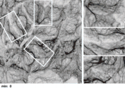
KFP-formation in the periphery of confluent
EK18-1 cells.
The confocal time lapse recording of 5 projected fluorescence images (inverse presentation; overview at left; high power views of boxed areas at right) reveals ongoing KFP formation close to the plasma membrane of tightly attached cells. The display rate is 15 frames/s, and images were taken every 90 seconds.
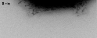
Microtubule-independent inward transport of KFPs
Fluorescence recording of HK18-YFP in a peripheral region of an EK18-1 cell treated with 100 µM nocodazole reveals continuing inward movement of newly-formed KFPs. Images were acquired every 20 s and the inhibitor was added at 18 min. The display rate is 10 frames/s.
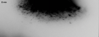
Need of intact actin filaments for inward-directed transport of newly synthesized keratin particles
Time-lapse movie of the cell edge of HK18-YFP fluorescence in an EK18-1 cell treated with the actin-polymerisation inhibitor cytochalasin D (1 µM). Images were taken every 20 s and the inhibitor was added at 18 min. Note the abrupt halt of KFP transport and the accumulation of KFPs in the cell periphery upon drug administration. The display rate is 10 frames/s.
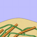
Animated model of keratin filament precursor formation and transport
The movie schematically shows the peripheral cytoplasm with an extending lamellipodium and depicts successive stages of cytoskeletal rearrangements in relation to focal adhesion formation. Initially, the lamellipodial cell edge is driven outward by cortical actin polymerization. Focal adhesion formation and maturation ensues followed by actin stress fibre anchorage. Then, KFPs appear at or near focal adhesion sites. Keratin particles enlarge and translocate along actin stress fibres toward the cell interior until integration into the peripheral KF network. For further details see Discussion. Microtubules are depicted in yellow. The display rate is 20 frames/s.