The keratin-filament cycle of assembly and disassembly
Kölsch A, Windoffer R, Würflinger T, Aach T, Leube RE
Continuous and regulated remodelling of the cytoskeleton is crucial for many basic cell functions. In contrast to actin filaments and microtubules it is not understood how this is accomplished for the third major cytoskeletal filament system consisting of intermediate filament polypeptides. Using time-lapse fluorescence microscopy of living interphase cells, in combination with photobleaching, photoactivation and quantitative fluorescence measurements, we observe that epithelial keratin intermediate filaments constantly release non-filamentous subunits, which are reutilized in the cell periphery for filament assembly. This cycle is independent of protein biosynthesis.
The different stages of the cycle occur in defined cellular subdomains: assembly takes place in the cell periphery, newly formed filaments are constantly transported toward the perinuclear region while disassembly occurs giving rise to diffusible subunits for another round of peripheral assembly. Remaining juxtanuclear filaments stabilize and encage the nucleus. Our data suggest that the keratin filament cycle of assembly and disassembly is a major mechanism of intermediate filament network plasticity allowing rapid adaptation to specific requirements, notably in migrating cells.
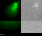
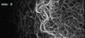
KFP appearance in the leading edge of migrating cells
Tableau of time-lapse recordings of HK18-YFP fluorescence (left) and corresponding phase contrast (right) of a migrating EK18-1 cell displaying multiple emerging KFPs in the proceeding lamellipodium. The bottom panel shows high power views of part of the lamellipodium. In this instance, cellular movement was compensated for with an image intensity-based method. The images were acquired every 30 s and are displayed at 30 frames/s.
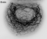
Persistence of KF network formation in the presence of the protein biosynthesis inhibitor cycloheximide
Time-lapse fluorescence microscopy of a PK18-5 cell treated with the protein biosynthesis inhibitor cycloheximide (17 µM). Images were acquired every 20 s and are displayed at 30 frames/s. The inhibitor was added after recording of 50 pictures. Note the continuous formation of new precursors.
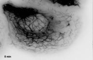
Persistence of KF network formation in the presence of the protein biosynthesis inhibitor puromycin
Time-lapse fluorescence recording of a PK18-5 cell treated with the protein biosynthesis inhibitor puromycin (1 µg/ml). Images were taken every 20 s and are displayed at 30 frames/s. The inhibitor was added after recording of 50 pictures. Note the continuous formation of new KFPs in the cell periphery.
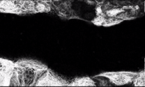
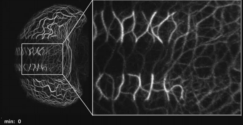
Detection of continuous inward-directed KF network motility
Time-lapse fluorescence recording of HK18-YFP in a section of a PK18-5 cell (projected images of 22 focal planes). Recording intervals were 60 s (display rate 25 frames/s). The frames are aligned to the first frame to compensate for cell movement. Note the continuous inward movement of KFs within the network.
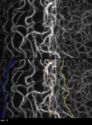
Detection of inward-directed KF network motility and loss of KFs by ROI-tracking
Time-lapse fluorescence recording of a HK18-YFP-producing PK18-5 cell after bleaching of three centripetal segments. Shown are projections of 25 focal planes, recording intervals were 2 min (display rate 50 frames/s). Note the inward movement of unbleached KF bundles and continuous loss of fluorescence without fragmentation, which are best seen at higher magnification at right.
Detection of inward-directed KF network motility
and loss of KFs by ROI-tracking
Time-lapse fluorescence recording of HK18-YFP in a segment of a PK18-5 cell (periphery at left, nucleus at right). Projection views of 11 focal planes are shown for each time point (recording intervals 60 s; 30 frames/s). ROIs demarcated in the lower panel were defined manually and the respective margins are shown by coloured lines. Note the continuous inward-directed flow and loss of fluorescent filaments during inward translocation of each ROI.
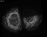
Detection of a continuous KF network turnover cycle by FRAP
Time-lapse fluorescence recordings were prepared from two PK18-5 cells producing HK18-YFP after bleaching of half of one of the cells. The movie presents a series of projected images (10 focal planes) after registration at a display rate 25 frames/s. Note the peripheral recovery of fluorescence and the continuous centripetal motility of the keratin system. Image stacks were recorded every 5 min.