Dissection of keratin network formation, turnover and reorganization in living murine embryos
Schwarz N, Windoffer R, Magin TM, Leube RE, 2015.
Epithelial functions are fundamentally determined by cytoskeletal keratin network organization. However, our understanding of keratin network plasticity is only based on analyses of cultured cells overexpressing fluorescently tagged keratins. In order to learn how keratin network organization is affected by various signals in functional epithelial tissues in vivo, we generated a knock-in mouse that produces fluorescence-tagged keratin 8. Homozygous keratin 8-YFP knock-in mice develop normally and show the expected expression of the fluorescent keratin network both in fixed and in vital tissues. In developing embryos, we observe for the first time de novo keratin network biogenesis in close proximity to desmosomal adhesion sites, keratin turnover in interphase cells and keratin rearrangements in dividing cells at subcellular resolution during formation of the first epithelial tissue. This mouse model will help to further dissect keratin network dynamics in its native tissue context during physiological and also pathological events.
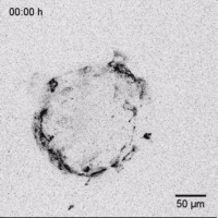
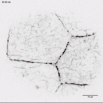
Time-lapse fluorescence recording (15-minute recording intervals) of an embryoid body derived from ESC clone 72 containing a Krt8-YFPneo allele. Note that only the outer cell layer corresponding to the primitive endoderm shows filamentous fluorescence in the cytoplasm. Some of these highly dynamic cells move outward upon attachment of the embryoid body to the gelatin-coated glass bottom dish. Scale bar, 50 μm.
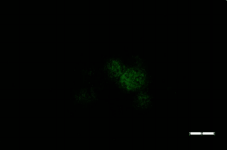

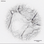
Animation of 3D-reconstruction depicts the Krt8-YFP fluorescence in the outer trophectoderm of a late blastocyst. Note the punctate accumulation at the plasma membranes in association with the developing cytoplasmic keratin network. The relative luorescence intensity is color coded. Scale bar, 20 μm.
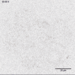
Time-lapse imaging of Krt8-YFP fluorescence in a developing homozygous knock-in 8-cell Embryo Recording intervals, 30 minutes. Note that diffuse cytoplasmic luorescence starts to appear around compaction (7 h) and that granular structures emerge subsequently at cell-cell borders. Scale bar, 20 μm.
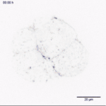
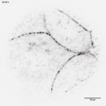
Time-lapse fluorescence microscopy of compacted homozygous Krt8-YFP morula at the transition to the blastocyst stage. Recording intervals, 15 minutes. The movie depicts a continuous increase of fluorescent puncta at cell-cell contact regions and highly motile filamentous structures in the cytoplasm. Scale bar, 20 μm.
Time-lapse recording of Krt8-YFP fluorescence in a mid-blastocyst (corresponding Figure 5f-f''; recording intervals, 210 seconds). The highly flexible cytoplasmic particles move throughout the cytoplasm at random. They fuse occasionally but also separate into smaller fragments. In contrast, the dotted cell border-restricted fluorescence is rather stable. Scale bar, 10 μm.
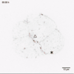
Time-lapse recording of Krt8-YFP fluorescence from the compacted morula to the blastocyst stage highlighting keratin reorganization during cell division. The first part of the video is identical to Supplementary Movie 6. Note the transient increase in diffuse fluorescence and the fragmented appearance of the network during pro-metaphase and the rapid formation of an extended filament network during anatelophase. Mitotic cells are labeled with triangle and asterisk. Scale bar, 10 μm.