The keratin-desmosome scaffold: pivotal role of desmosomes for keratin network morphogenesis.
Moch M, Schwarz N, Windoffer R, Leube RE, 2019
Desmosome-anchored keratin intermediate filaments (KFs) are essential for epithelial coherence. Yet, desmosomal KF attachment and network organization are still unexplored in vivo. We, therefore, monitored KF network morphogenesis in fluorescent keratin 8 knock-in murine embryos revealing keratin enrichment at newly formed desmosomes followed by KF formation, KF elongation and KF fusion. To examine details of this process and its coupling to desmosome formation, we studied fluorescent keratin and desmosomal protein reporter dynamics in the periphery of expanding HaCaT keratinocyte colonies. Less than 3 min after the start of desmosomal proteins clustering non-filamentous keratin enriched at these sites followed by KF formation and elongation. Subsequently, desmosome-anchored KFs merged into stable bundles generating a rim-and-spokes system consisting of subcortical KFs connecting desmosomes to each other and radial KFs connecting desmosomes to the cytoplasmic KF network. We conclude that desmosomes are organizing centers for the KF cytoskeleton with a hitherto unknown nucleation capacity.
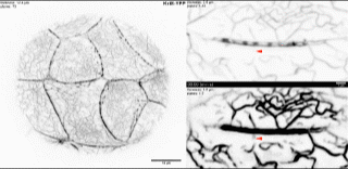
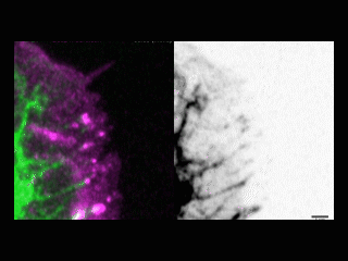
Keratin filaments nucleate close to the cortical interdesmosomal network in murine blastocysts. The overview at left shows an animated maximum intensity projection of the fluorescence recorded in the trophectoderm of a late murine Krt8-YFP knock-in blastocyst (see also corresponding Fig. 1c). The time-lapse fluorescence images at right were recorded at the cell border of two trophectoderm cells of the same blastocyst every 62.5 s (see also corresponding Fig. 1d). The top reveals the appearance of growing keratin particles from the interdesmosomal subcortical keratin system. These motile and highly flexible particles fuse and enlarge before merging with the cytoplasmic KF network (arrowheads delineate a few selected examples; contrast was enhanced per frame to compensate for bleaching). The lower plane shows the same region at enhanced contrast settings to unravel further details in the selected focal planes.

The 3D animation of a z-stack recording depicts the fluorescence of keratin and desmosome reporters in a confluent HaCaT cell culture. The images (maximum intensity projection) were obtained from mixed HaCaT cell cultures containing cells producing HK5-EYFP (clone B10) delineating KFs and Dsc2-mCerulean delineating desmosomes (for details see legend to corresponding Fig. 2b). The animation first shows rotating views of the keratin-desmosome fluorescence, then rotating views of the desmosome fluorescence by itself, and then top-to-bottom views of single focal planes of double keratin-desmosome fluorescence.

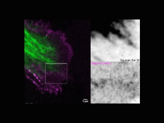
Scroll through confocal sections reveals organizational details of the keratin-desmosome scaffold in a confluent HaCaT cell culture. The images were obtained from HaCaT cells producing HK5-EYFP delineating KFs and Dsc2-mCerulean delineating desmosomes (for details see legend to corresponding Fig. 2b). Note the presence of desmosome-anchored interdesmosomal and radial KFs in different focal planes.
Time-lapse image series depicts the coordinated dynamics of HK5-YFP and Dsc2-mCerulean in a confluent HaCaT culture. The images (maximum intensity projections) were recorded every 55 s in HaCaT cells producing HK5-EYFP delineating KFs and Dsc2-mCerulean delineating desmosomes (for details see legend to corresponding Fig. 2b). Note the coordinated movement of desmosomes with attached subcortical and radial KFs.
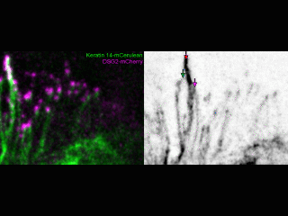
The time-lapse image series depicts the coordinated dynamics of keratin 14-mCerulean and DspI-mApple in a confluent HaCaT cell culture. The images (maximum intensity projections; 45 s recording intervals) were obtained from HaCaT cells producing Keratin 14-mCerulean and DspI-mApple delineating KFs and desmosomes (for details see legend to corresponding Fig. 2c). Note the coordinated movement of desmosomes with attached subcortical and radial KFs.
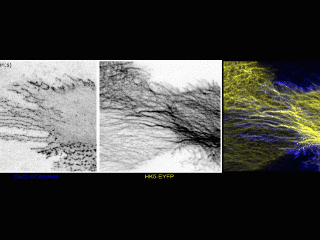
Elongating keratin filaments nucleate at nascent desmoplakin clusters at the outermost cell border of an expanding HaCaT colony. The time-lapse series (maximum intensity projections; 15 s recording intervals) shows DspI-mApple and K14-mCerulean in two adjacent HaCaT cells. Examples of clustering DspI-mApple are delineated by magenta arrows, the ends of elongating KFs by green arrows. For further details see legend to corresponding Fig. 3.
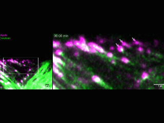
Elongating keratin filaments nucleate at nascent desmocollin 2 clusters. The time-lapse series (maximum intensity projections; 26 s recording intervals) shows the fluorescence of Dsc2-mCerulean and HK5-EYFP at the expanding cell-cell border of a HaCaT cell next to a non-transfected HaCaT cell in the periphery of an expanding HaCaT colony. Examples of clustering Dsc2-mCerulean are delineated by magenta arrows, the ends of elongating KFs by green arrows. For further details see legend to corresponding Fig. 4a.
Elongating keratin filaments nucleate at nascent desmoglein 2 clusters. The time-lapse series (maximum intensity projections; 26 s recording intervals) shows the fluorescence of Dsg2-mCherry and Keratin 14-mCerulean at the expanding cell-cell border of a HaCaT cell next to a non-transfected HaCaT cell in the periphery of an expanding HaCaT colony. Examples of clustering Dsg2-mCherry are delineated by magenta arrows. For further details see legend to corresponding Fig. 4d.
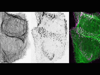
Subcortical, interdesmosomal keratin filaments merge into bundles. The time-lapse series (maximum intensity projections; 60 s recording intervals) shows Keratin 14-mCerulean and Dsg2-mCherry fluorescence at the cell border in a HaCaT cell colony 3 days after seeding (see also corresponding Fig. 5a). Note the fusion of color-coded interdesmosomal KFs into KF bundles with tightly-spaced Dsg2-mCherry-positive desmosomes producing a pearls-on-a-string pattern.
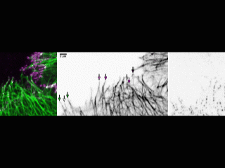
Interdesmosomal and radial keratin filaments merge into bundles. The time-lapse series (maximum intensity projections; 26 s recording intervals) shows HK5-EYFP and Dsc2-mCerulean fluorescence at the cell border in a HaCaT cell colony 3 days after seeding (see also corresponding Fig. 5b). Note the fusion of color-coded interdesmosomal and radial KFs into KF bundles with tightly-spaced and enlarged Dsc2-mCerulean-positive desmosomes.