Keratin intermediate filaments: intermediaries of epithelial cell migration
Yoon S, Leube RE, 2019
Migration of epithelial cells is fundamental to multiple developmental processes, epithelial tissue morphogenesis and maintenance, wound healing and metastasis. While migrating epithelial cells utilize the basic acto-myosin based machinery as do other non-epithelial cells they are distinguished by their copious keratin intermediate filament cytoskeleton, which comprises differentially expressed members of two large multigene families and presents highly complex patterns of posttranslational modification. We will discuss how the unique mechanophysical and biochemical properties conferred by the different keratin isotypes and their modifications serve as finely-tunable modulators of epithelial cell migration. We will furthermore argue that KFs together with their associated desmosomal cell-cell junctions and hemidesmosomal cell-extracellular matrix adhesions serve as important counterbalances to the contractile acto-myosin apparatus either allowing and optimizing directed cell migration or preventing it. The differential keratin expression in leaders and followers of collectively migrating epithelial cell sheets provides a compelling example of isotype-specific keratin functions. Taken together, we conclude that the expression levels and specific combination of keratins impinge on cell migration by conferring biomechanical properties on any given epithelial cell affecting cytoplasmic viscoelasticity and adhesion to neighboring cells and the extracellular matrix.
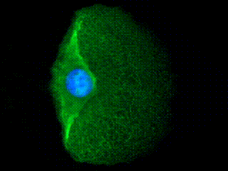
3D animation of a human primary keratinocyte stained with anti-pan-keratin antibody (green) and phalloidin binding to actin (red) is shown. Note the distinct distribution patterns of the two cytoskeletal networks where KFs form a much more bundled network around the nucleus and are distributed throughout the entire height of the cell, whereas actin filaments form a flat network at the base. Scale bar: 20 μmperinuclear cage. Scale bar: 10 μm