combines expertise in macroscopic and microscopic anatomy. It is dedicated to interdisciplinary approaches in teaching and research. MOCA is a major partner in the innovative medical curriculum.
Lectures and laboratory instruction in the areas of developmental, cell and molecular biology as well as in all areas of histology and gross anatomy are part of its general educational commitment in the life sciences. As lead beneficiary MOCA coordinates the Research Training Group MEƎT, which is funded by the German Research Council (DFG).
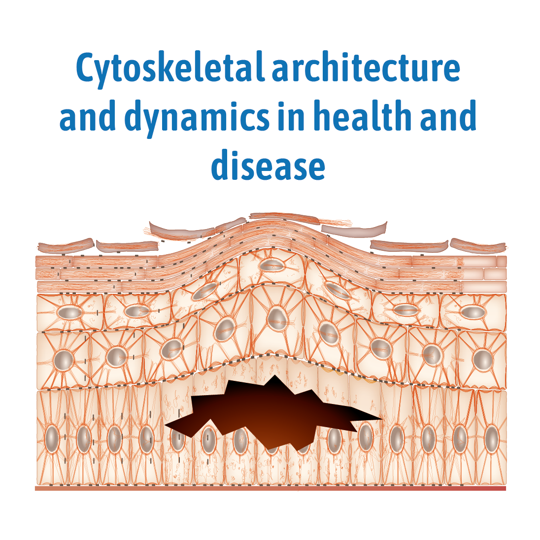
The cytoskeleton consists of interconnected multimeric filaments, which physically link subcellular compartments and connect cells with their neighbors and the surrounding extracellular matrix. The traditional view that the cytoskeleton serves a primary mechanical role by providing a highly plastic but also resilient scaffold is expanding by the realization that it is intricately involved in all cellular processes and thereby fulfils fundamental functions in maintaining homeostasis at the cellular, tissue and organismal level.
Our research is driven by the tenet that structure determines function. We therefore employ multidimensional and multimodal microscopic methods to study structure-function relationships at different time and length scales in cultured cells and living organisms. Main emphasis is on the intermediate filament cytoskeleton, which displays unique properties in comparison to the actin filament- and microtubule-based cytoskeletal networks. Its rod-shaped polypeptide subunits assemble spontaneously in the absence of co-factors into highly extensible and flexible filaments (see Figure 1) and filament bundles, which are attached to desmosomes at cell-cell contact sites and to hemidesmosomes at cell-extracellular matrix interfaces in epithelial cells. The compositional diversity of intermediate filaments reflects their high degree of functional adaptation to cell type- and context-dependent properties.
MOCA hosts several working groups, which are united by their common interest in the properties of the cytoskeleton within cells, its coordination within tissues and in relation to the extracellular matrix and its relevance for cell and tissue function. MOCA has been involved in the organization of graduate and postgraduate programs (Marie Skłodowska-Curie Innovative
Training Network on Integrated Component Cycling in Epithelial Cell Motility [InCeM], https://incem.rwth-aachen.de/; Isaac Newton Institute for Mathematical Sciences Programme on Coupling geometric PDEs with physics for cell morphology, motility and pattern formation, and is currently administrating the DFG graduate school Mechanobiology in Epithelial 3D Tissue Constructs.
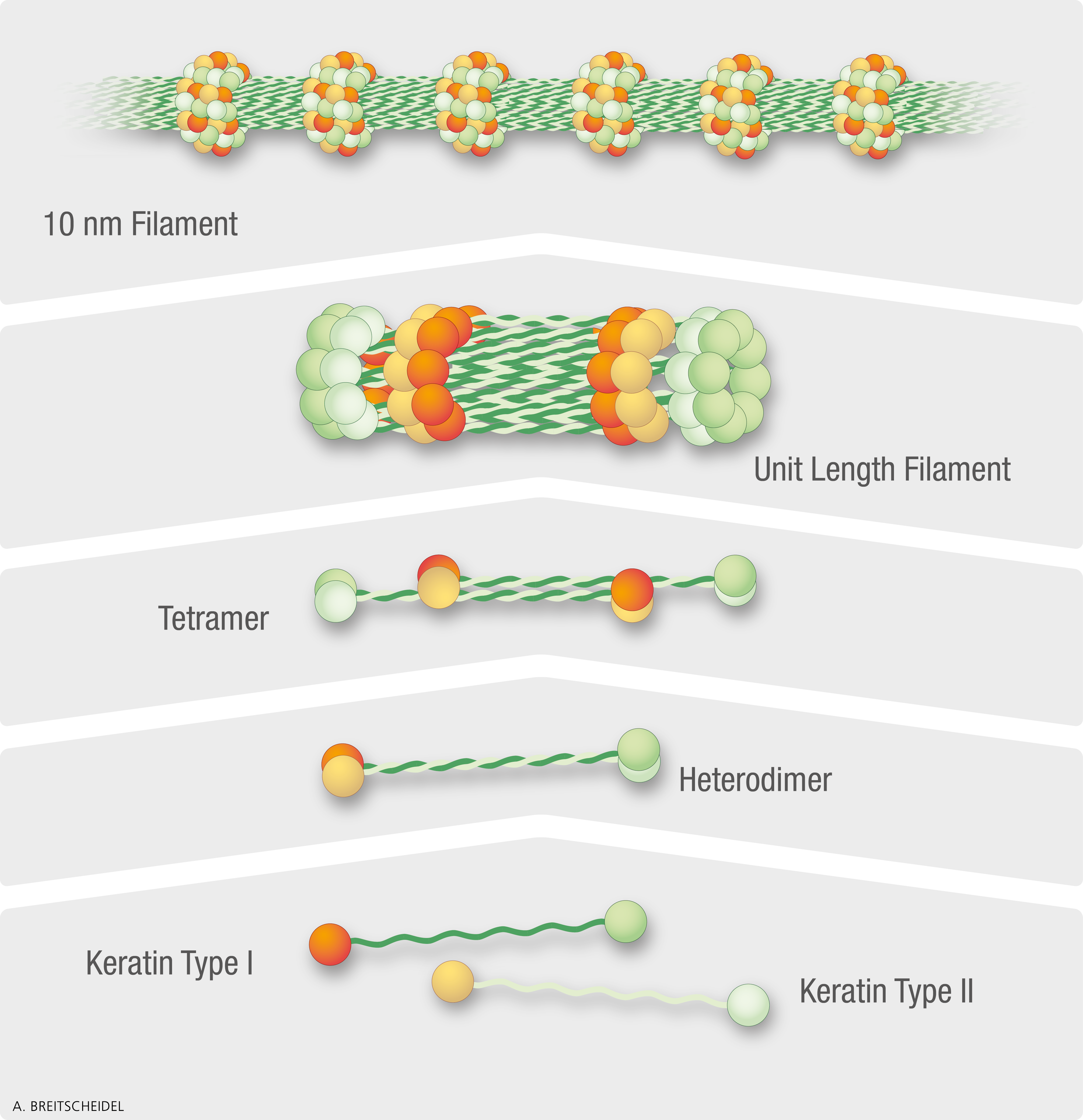
Scheme depicting aspects of cofactor-independent keratin filament assembly.
Keratins are grouped into type I and type II. Each keratin polypeptide consists of an ∼310 amino acid-long central helical rod domain that is flanked by variable head and tail domains. Dimers form by coiled-coil interactions between the rod domains of a type I and type II keratin polypeptide. The resulting heterodimers subsequently assemble in an antiparallel and staggered fashion into non-polar tetramers. Typically, four to eight keratin tetramers associate laterally to generate the ∼65 nm unit length filament. Unit length filaments elongate by end-on intercalation into 10-nm filaments. (The figure is taken from Yoon and Leube, 2019, Essays Biochem 63:521)