Florian Geisler and Rudolf Leube with Jadirah Sarmad
To dissect the molecular mechanisms of cytoplasmic intermediate network morphogenesis and its function in a living organism we use the genetic model organism Caenorhabditis elegans. Of particular interest is the intestinal intermediate filament cytoskeleton, which is composed of six different intermediate filament polypeptides. It is localized below the adluminal terminal web region and is anchored to the apical junction complex. This arrangement is conserved from the nematode to humans.
In genetic screens we have identified structural and enzymatic regulators of intestinal intermediate filament network formation. By targeted depletion of intermediate filament polypeptides we have furthermore identified isotype-specific consequences on network formation. The different mutations elicit distinct morphological phenotypes in the intestine that can be correlated with different dysfunction affecting biomechanics, signaling and metabolism with consequences on time of development, body size, brood size, survival and overall stress resilience.
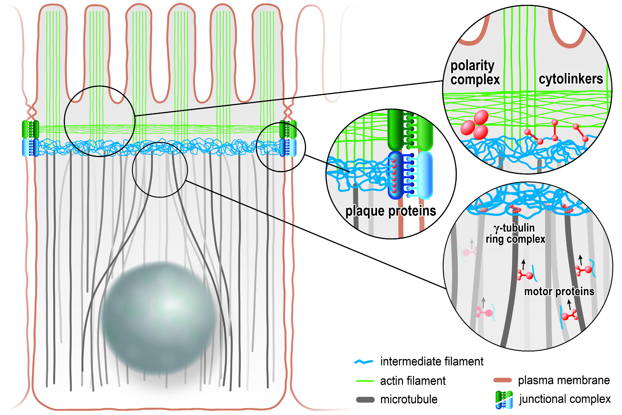
Figure 1. The scheme summarizes the integration of the intermediate filament network into the apical cytoskeleton of the C. elegans intestine (taken from Coch and Leube, 2016, Cells 5:32).