RESEARCH      PUBLICATIONS      MOVIE GALLERY      TEAM      CONTACT

Keratin filaments mediate the expansion of extra-embryonic membranes in the post-gastrulation mouse embryo

Nahaboo W , Eski SE , Despin-Guitard E, Vermeersch M, Versaevel M , Saykali B, Monteyne D, Gabriele S, Magin TM, Schwarz N , Leube RE, Zwijsen A, Perez-Morga D, Singh SP, Migeotte I

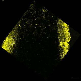
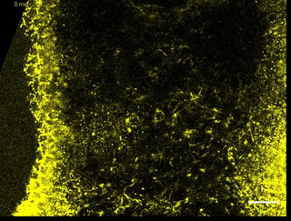
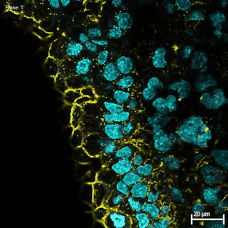
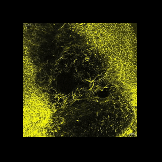
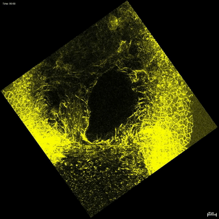
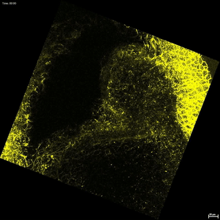
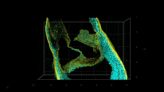
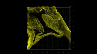
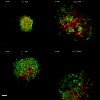
Mesoderm arises at gastrulation and contributes to both the mouse embryo proper and its extra-embryonic membranes. Two-photon live imaging of embryos bearing a keratin reporter allowed recording filament nucleation and elongation in the extra-embryonic region. Upon separation of amniotic and exocoelomic cavities, keratin 8 formed apical cables co-aligned across multiple cells in the amnion, allantois, and blood islands. An influence of substrate rigidity and composition on cell behavior and keratin content was observed in mesoderm explants. Embryos lacking all keratin filaments displayed a deflated extra-embryonic cavity, a narrow thick amnion, and a short allantois. Single-cell RNA sequencing of sorted mesoderm cells and micro-dissected amnion, chorion, and allantois, provided an atlas of transcriptomes with germ layer and regional information. It defined the cytoskeleton and adhesion expression profile of mesoderm-derived keratin 8-enriched cells lining the exocoelomic cavity. Those findings indicate a novel role for keratin filaments in the expansion of extra-embryonic structures and suggest mechanisms of mesoderm adaptation to the environment.

3D reconstruction in the ExE region of a fixed whole-mount embryo stained for F-actin (Phalloidin) (n=4). Z interval is 1 µm.


Download: .mp4 / .wmv

Z-projection from two-photon live imaging of one K8-eYFP (yellow) LS embryos, when YFP becomes detectable in ExE mesoderm (n=9). Time interval is 15 min, scale bar represents 25 µm.


Download: .mp4 / .wmv

Z-projection from two-photon live imaging of a K8-eYFP (yellow) 0B embryos at 40X magnification showing whole ExE region (n=10). Time interval is 15 min, scale bar represents 25 µm.


Download: .mp4 / .wmv

Z-stack of a whole-mount fixed K8-eYFP (yellow) LB embryo stained for nuclei (DAPI, cyan) focused on ExE region (n=15). Z interval is 1 µm, scale bar represents 20 µm.


Download: .mp4 / .wmv

Z-projection from two-photon live imaging of 5 K8-eYFP (yellow) EB/LB embryos focusing on ExE region at 40X magnification highlighting K8 containing cables (n=10). Time interval is 15 min and scale bars represent 25 µm.


Download: .mp4 / .wmv

Z-projection from two-photon live imaging of 2 K8-eYFP (yellow) LB embryos focusing on ExE region at 40X magnification and showing the amnio-chorionic fold at the anterior separation point (n=6). Time interval is 15 min and scale bars represent 25 µm.


Download: .mp4 / .wmv

Z-projection from two-photon live imaging of 3 K8-eYFP (yellow) LB embryos focusing on ExE region at 40X magnification showing allantois in lateral (a, b) (n=12) and posterior (c) views (n=3). Time interval is 15 min and scale bars represent 25 µm..


Download: .mp4 / .wmv

First example of 3D reconstructions in the ExE region of 2 fixed whole-mount K8-eYFP (yellow) embryos stained with DAPI (cyan) (n=15). Z interval is 1 µm.


Download: .mp4 / .wmv

Second example of 3D reconstructions in the ExE region of 2 fixed whole-mount K8-eYFP (yellow) embryos stained with DAPI (cyan) (n=15). Z interval is 1 µm.


Download: .mp4 / .wmv

Z-projections of E (left) and ExE (right) mesoderm explants seeded on a hard substrate coated with Fn1 (top) and ColIV (bottom). The time interval is 15 min (ColIV coating) and 20 min (Fn1 coating) and the scale bar represents 100 µm


Download: .mp4 / .wmv