Visualization of gap junction mobility in living cells
Windoffer R, Beile B, Leibold A, Thomas S, Wilhelm U, Leube RE, 2000
In order to study the dynamics of gap junctions in living cells, a cDNA was expressed in hepatocellular carcinoma-derived PLC cells coding for chimerical polypeptide Cx.EGFP-1, which consists of rat connexin32 and enhanced green fluorescent protein (EGFP). Cx.EGFP-1 was integrated into gap junctions, and the emitted epifluorescence reliably reported the distribution of the chimera. Therefore, stably transfected PLC clone PCx-9 was used to examine the dynamic behavior of gap junctions by time-lapse fluorescence microscopy.
The pleomorphic fluorescent junctional plaques were highly motile within the plasma membrane. They often fused with each other or segregated into smaller patches, and fluctuation of fluorescence was detected within individual gap junctions. Furthermore, the uptake of junctional fragments into the cytoplasm of live cells was documented as originating from dynamic invaginations that form long tubulovesicular structures that pinch off. Endocytosis and subsequent lysosomal degradation, however, appeared to contribute only a little to the rapid gap junction turnover (determined half-life of 3.3 h for Cx.EGFP-1), since most cytoplasmic Cx.EGFP-1 fluorescence did not colocalize with the endocytosed fluid phase marker horseradish peroxidase or the receptor-specific endocytotic ligand transferrin and since it was distinct from lysosomes.
Disassembly of gap junctions was monitored in the presence of the translation-inhibitor cycloheximide and showed increased endocytosis and continuous reduction of junctional plaques. Highly motile cytoplasmic microvesicles, which were detectable as multiple, weakly fluorescent puncta in all movies, are proposed to contribute significantly to gap junction morphogenesis by the transport of small subunits between biosynthetic, degradative, and recycling compartments.
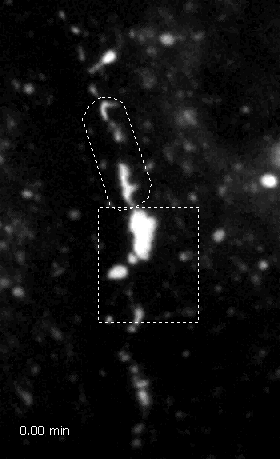
Time-lapse fluorescence microscopy documenting dynamics of Cx.EGFP-1-positive structures in living PLC cells of clone PCx-9. Note the different types of mobility of fluorescent patches at cell contact sites. A region of high mobility is labeled by rectangle, fusion of plaques is demarcated by ovoid between 0.00 min and 9.50 min and again in another area by ovoid between 9.75 min and 28 min. Separation of a plaque into several fragments is marked by circle. The time points of recording are given at lower left corner in min. Some strongly fluorescent, large structures that remain stationary are seen in the cytoplasm. Note also the presence of many rapidly moving, small cytoplasmic dots. Pictures taken every 15 seconds for 28 minutes.
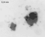
Fluorescence micrographs (inverse presentation) depicting dynamic heterogeneities in two Cx.EGFP-1-positive cell contact sites that are viewed en face in live PLC cells of clone PCx-9. The time of recording is given in the upper left corner. Note the high degree of mobility of patches with differing fluorescence intensity within the gap junctions shown. In addition, weak and highly mobile fluorescence is also detectable in the vicinity corresponding to cytoplasmic regions. Pictures taken every 8.4 seconds for 9.94 minutes.
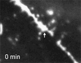
Time-lapse fluorescence microscopy of Cx.EGFP-1-expressing PCx-9 cells depicting endocytosis of gap junctional fragments. A strongly fluorescent patch (arrow) can be tracked within a gap junction which first extends into the cytoplasm (9.5 min), retracts (20.5 min) before it eventually buds off (22.5 min) and is taken up into the cytoplasm (27 min). Pictures taken every 30 seconds for 27 minutes.
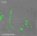
Series of overlay pictures showing brightfield pictures merged with fluorescence pictures (green color) to demonstrate Cx.EGFP-1 mobility in four PCx-9 cells before and after addition of cycloheximide (17.1 µM). The cells remained viable during the entire period of 249 min as judged from the ongoing undulations of peripheral cell regions and the continuous fast movements of cytoplasmic dots. Note the stationary nature of large cytoplasmic vesicular structures contrasting with the highly dynamic plaques at cell contact sites which are subject to continuous remodeling. Between 199 min and 204 min, i.e., almost 3 h after addition of cycloheximide a large plaque is broken up (upper left). Concurrently, a cell slides between the two cells establishing new contacts. Note the overall reduction in fluorescent structures during cycloheximide treatment, most notably at cell contact sites. Pictures taken every 60 seconds for 249 minutes.