Identification of novel principles of keratin filament network turnover in living cells
Windoffer R, Wöll S, Strnad P, Leube RE, 2004
It is generally assumed that turnover of the keratin filament system occurs by exchange of subunits along its entire length throughout the cytoplasm. We now present evidence that a circumscribed submembranous compartment is actually the main site for network replenishment. This conclusion is based on the following observations in living cells synthesizing fluorescent keratin polypeptides:
1) Small keratin granules originate in close proximity to the plasma membrane and move toward the cell center in a continuous motion while elongating into flexible rod-like fragments that fuse with each other and integrate into the peripheral KF network.
2) Recurrence of fluorescence after photobleaching is first seen in the cell periphery where keratin filaments are born that translocate subsequently as part of the network toward the cell center.
3) Partial keratin network reformation after orthovanadate-induced disruption is restricted to a distinct peripheral zone in which either keratin granules or keratin filaments are transiently formed.
These findings extend earlier investigations of mitotic cells in which de novo keratin network formation was shown to originate from the cell cortex. Taken together, our results demonstrate that the keratin filament system is not homogeneous but is organized into temporally and spatially distinct subdomains. Furthermore, the cortical localization of the regulatory cues for keratin filament turnover provides an ideal way to adjust the epithelial cytoskeleton to dynamic cellular processes.
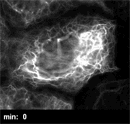
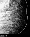
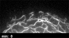
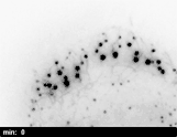
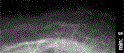
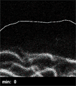
Time-lapse confocal laser scanning microscopy of a peripheral free region of a human primary keratinocyte depicting the inward-directed aviement of HK14-YFP fluorescence which originates in a submembraneous compartment and results in integration of rod-like structures into the peripheral filament network. Recording intervals: 60 s.
Time-lapse confocal laser scanning microscopy of a peripheral free region of a human primary keratinocyte depicting the inward-directed aviement of HK14-YFP fluorescence which originates in a submembraneous compartment and results in integration of rod-like structures into the peripheral filament network. Recording intervals: 60 s.