RESEARCH      PUBLICATIONS      MOVIE GALLERY      TEAM      CONTACT

Multidimensional Monitoring of Keratin Intermediate Filaments in Cultured Cells and Tissues

Schwarz N, Moch M, Windoffer R, Leube RE, 2016

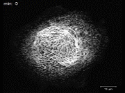
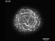
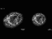
Keratin filaments are a hallmark of epithelial differentiation. Their cell type-specific spatial organization and dynamic properties reflect and support epithelial function. To study this interdependency, imaging of fluorescently tagged keratins is a widely used method by which the temporospatial organization and behavior of the keratin intermediate filament network can be analyzed in living cells. Here, we describe methods that have been adapted and optimized to dissect and quantify keratin intermediate filament network dynamics in vital cultured cells and functional tissues.

Time-lapse fluorescence recording of keratin 13-EGFP in AK13-pax1 cells. Major steps of the keratin cycle, namely nucleation, elongation, integration, and bundling, are seen.


Download: .mp4 / .wmv

Maximum intensity projections of keratin 13-EGFP fluorescence detecting keratin network reorganization during cell division.


Download: .mp4 / .wmv

Denoised recordings of an AK13-1 cell expressing keratin 13-EGFP in a nonnormalized (left) and normalized (right) format. Note that normalization leads to occasional jumps because of extension/shrinkage of the overall network (time points 13 and 14 min).


Download: .mp4 / .wmv

Time-lapse fluorescence recording of a compacted Krt8-YFP embryo shows the first appearance of keratins 10 h after compaction.


Download: .mp4 / .wmv

Animation of a 3D reconstruction of a Krt8-YFP blastocyst.


Download: .mp4 / .wmv